

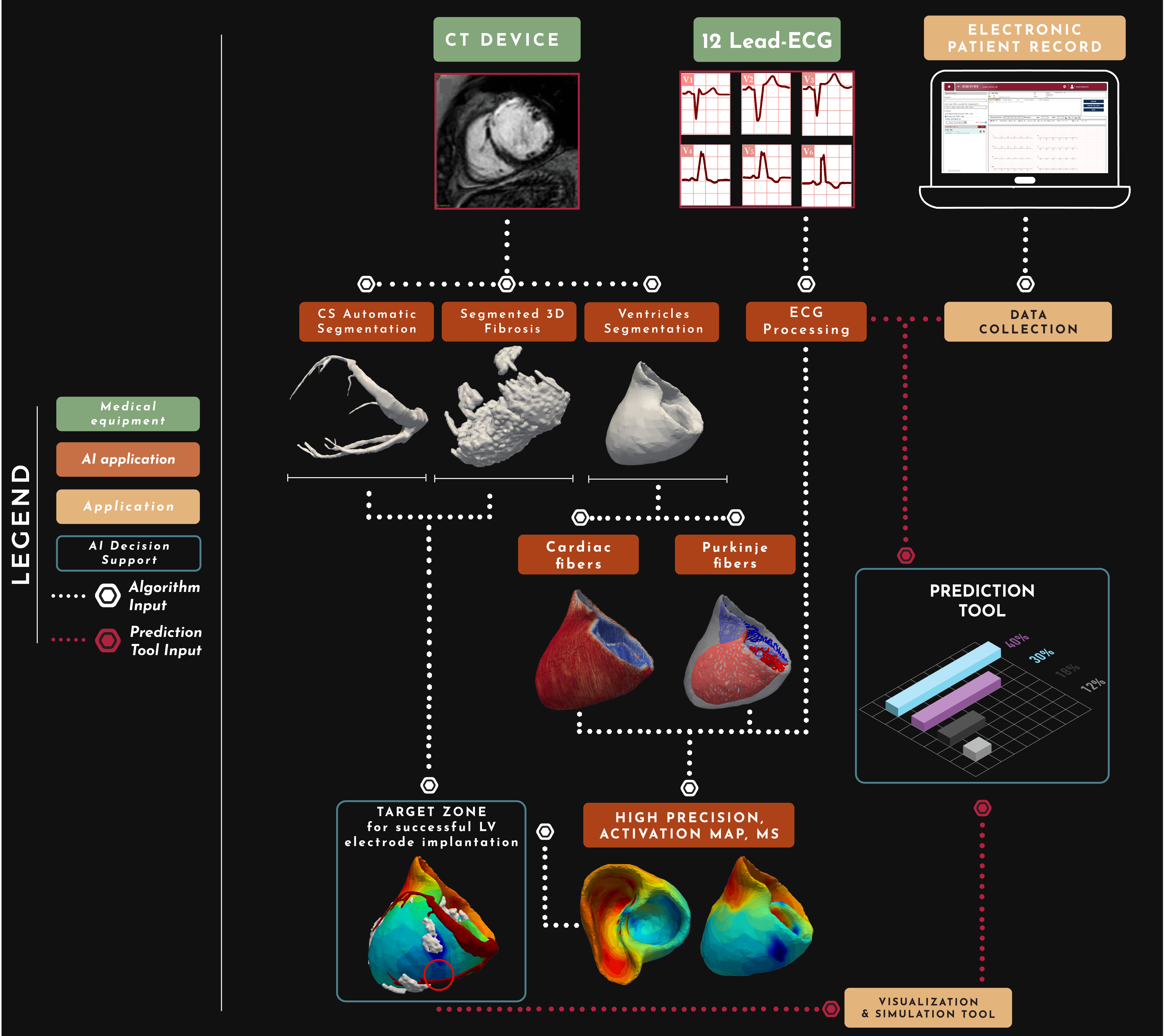
THE SOLUTION

OUR SOLUTION
What we predict
We developed a comprehensive tool for successful CRT implantation and preparation before the procedure. Prediction may help to select patients who most probably will be non-responders (based on their clinical characteristics) using standard approach.


VENTRICLES SEGMENTATION
Ensemble of three 2D neural networks, one for each anatomical plane
Automatic segmentation of ventricles
Three 2D neural networks analyse independently the three anatomical planes of the CT 3D image: the sagittal, axial and coronal plane. Those contributions are combined to create a segmentation space where the whole ventricle structure is identified and isolated from the image. A further post-processing procedure refines the output removing noise and filling small gaps and holes.
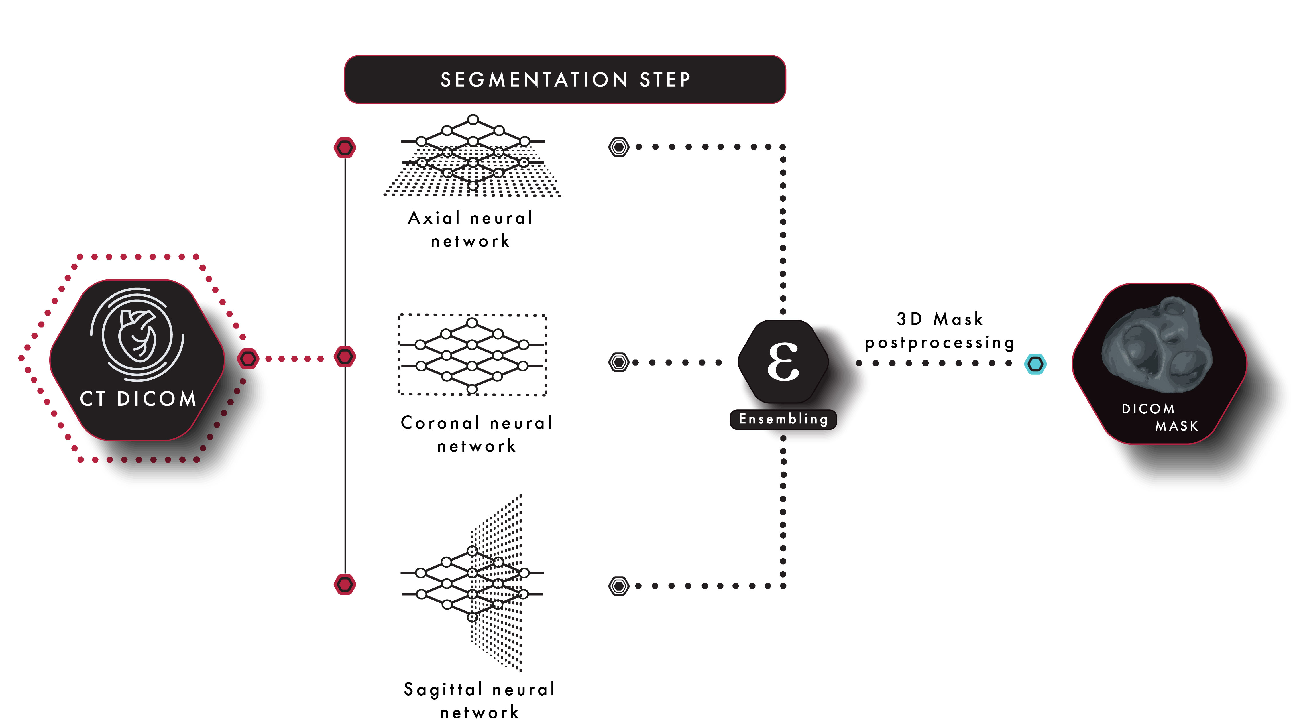
3D ventricles models
Results of segmentation
Mouse pointer: left = rotate, right= move (pan),
mouse wheel = zoom in/out
Mobile finger use to rotate and zoom in/out

CORONARY VEINS
SEGMENTATION
One algorithm and four neural networks optimize segmentation
of Coronary Veins along its branches

Automatic segmentation of coronary veins
The segmentation of coronary veins is a sophisticated task, due to its characteristic branching structure, thus requires a multistep pipeline:
An ensemble of three 2D neural networks analyzes independently the three anatomical planes of the CT 3D image, in the same fashion as for ventricles segmentation.
This preliminary segmentation is then processed by a patented vein-tracking algorithm which starts an iterative refinement cycle along the branches of the pre-segmented structure.
After multiple interactions with a specialized neural network, the final segmentation space is created.
A further post-processing procedure finalizes the output removing noise and filling small gaps and holes.
3D coronary veins models
Results of segmentation
Mouse pointer: left = rotate, right= move (pan),
mouse wheel = zoom in/out
Mobile finger use to rotate and zoom in/out
Ventricle & Veins overlay
Overlay
The final segmentations of the ventricles and coronary veins, shown together, give an accurate idea of the morphology of the patient’s heart and coronary veins before implantation.


LAO projection

RL projection

RAO projection
AP projection

We believe in healthcare visionaries.

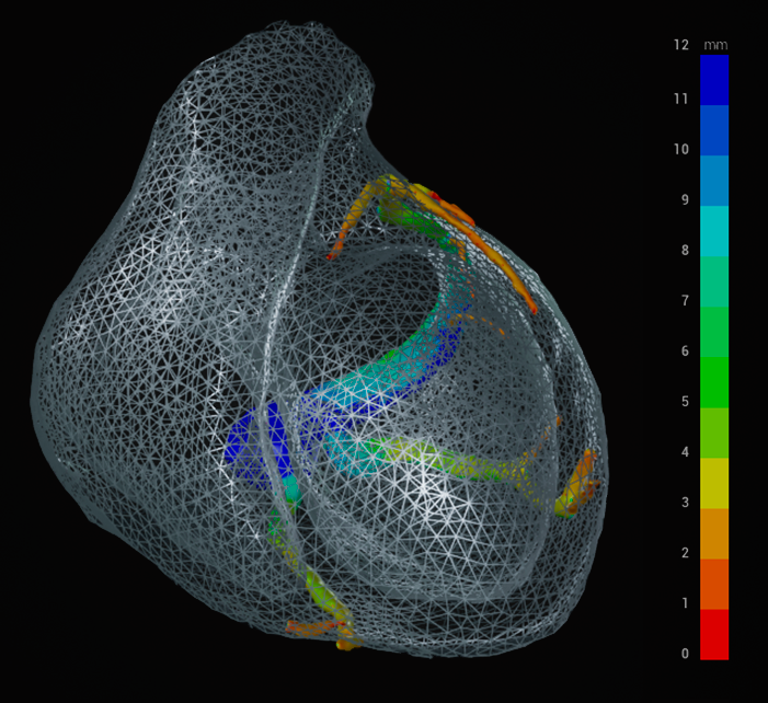
TARGET ZONE
Successful LV lead inplantation
Information and clear visualization of morphology of the heart and coronary veins, high precision 3D activation map and areas of fibrosis provide an unbeatable resource for an accurate identification of the target zone. This represents the target vein for left ventricular lead implantation that can yield the highest chance of success with CRT for that individual patient.

Grey: fibrosis
Red: coronary veins
3D SIMULATION TOOL
An immersive tool allows the cardiologist to have a synergistic view of the patient's anatomy, thanks to:
- The possibility to overlay the two 3D representations of coronary sinus and ventricles.
- The interactive interface which allows to zoom, pan, and select a projection plane.
- The power of choosing among multiple layers of information such as veins diameters, electric ventricles activation map, distance calculations and vein sections visualization.
- The accurate simulation tool that reproduces the access of a catheter in the coronary sinus yielding a forecast of the complexity and feasibility of the left ventricle lead implantation for the specific patient.
